Sidebone

Veterinary advice should be sought before applying any treatment or vaccine.

Sidebone

Sidebone is the ossification of the collateral cartilages of the foot. The process results in the normally soft cartilages found on the inside and outside aspects of the foot, to harden and become less flexible bone.
Sidebone Comparison
The condition occurs most commonly in Heavy draft, Cob, and Icelandic breeds, and horses with poorly balanced feet or uneven loading of the heels. Sidebone usually occurs in the forelimb, cases involving the hindlimb are usually associated with acute trauma or chronic repetitive trauma (such as punctures, chronic bruising from interference, or lacerations), resulting in chronic inflammatory changes and osseous metaplasia of the cartilage.

Diagnosis of Sidebone


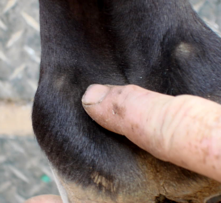
The lateral cartilages provide support and protection for the soft tissues at the back of the foot. Ossification typically starts from distal to proximal, and may initially induce lameness in affected horses. The only way to verify the extent of ossification that has occurred is through radiographic images. Nuclear scintigraphic examination may also be helpful to help differentiate between a fractured sidebone and a separate center of ossification.

Symptoms

Prominent, hard, rigid and bony enlargement proximal to the coronary band
Short and choppy stride
Uneven shoe wear
Lameness

Diagnosis

  • History
  • Clinical signs
  • Physical exam
  • Radiographs
  • Nuclear scintigraphic exam

Support

Therapies

TherapiesDetails
Regular hoof trimmingFeet should be kept properly balanced to distribute the weight-bearing evenly, conducted by an experienced farrier at regular intervals less than 6 weeks in duration.
Bar shoesMay be useful in the presence of lateromedial foot imbalance.

Prevention

  • Ensure horse gets shod in regular intervals.
  • Avoid lots of work on hard ground such as roads.

Scientific Research

General Overviews

  • sidebone icon
  • sidebone icon

Risk Factors

  • Heavy draft, Cob, and Icelandic breeds
  • Poor hoof conformation- especially horses with narrow, upright feet, pigeon-toed or splay-footed.
  • Frequent exercise on hard surfaces
  • Previous over reaching injury or trauma to the cartilages from wire cuts or other injury that causes direct damage to the cartilages.
  • Concurrent collateral ligament desmitis of the DIP joint
  • Horses with a low height to bodyweight ratio
  • Abnormal limb conformation, including angular limb deformities, which cause uneven forces on the hoof and collateral cartilages.