Snapper is a 22-year old Missouri Fox Trotter gelding who has been Bridget’s horse almost her entire life—she even saw him being born on her family’s farm. He was Bridget’s best friend and constant companion while she was growing up. Being a gaited horse, Snapper and Bridget mainly competed in breed specific horse shows, although covering a variety of disciplines—including trail, jumping, barrel racing, and gaited pleasure classes. As Bridget and Snapper both grew older, they stopped showing and instead enjoyed pleasure riding on trails with friends. He was also a great lesson horse for younger riders when Bridget taught riding lessons.
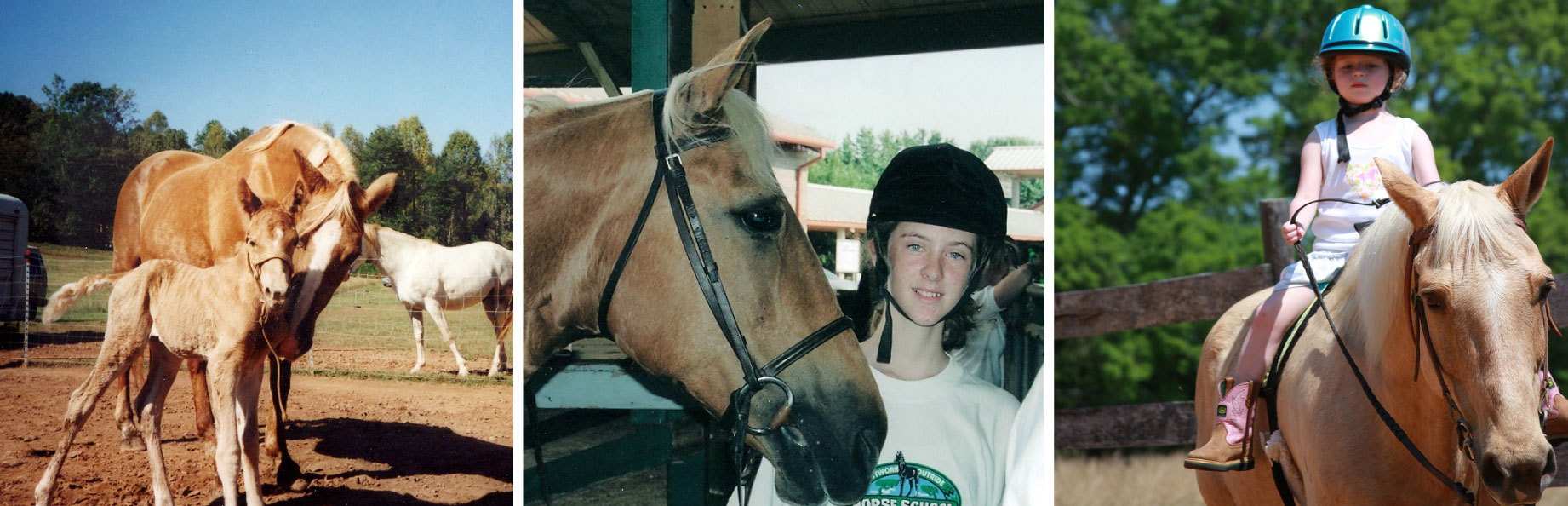
Snapper as a foal (left), competing w/ Bridget (middle), and as a lesson horse (right)
Seeking an Explanation
June of 2013, Snapper turned up lame in his right forelimb. Bridget discovered that he had developed a foot abscess in his right front foot. Bridget treated it with stall rest and frequent Epsom salt soakings until it healed. Over the next year, Snapper developed 4 more abscesses in the same location, in the same foot. Each time, Bridget called a local veterinarian out to look at the foot, who would diagnosis it as an abscess. One time radiographs were taken of Snapper’s foot, and the vet still diagnosed it as simply an abscess.
Bridget was beyond frustrated, because she knew that there had to be a reason Snapper was getting so many abscesses, however none of the local vets were able to explain why. The usual response she got was that “it’s been an unusually bad year for abscesses.” She decided to seek expertise elsewhere---and made an appointment for Snapper at the University of Georgia’s (UGA) Large Animal Hospital.
First Vet Visit at UGA
Upon arrival at UGA, Bridget and Snapper were promptly greeted by one of the veterinary interns who started asking questions about Snapper’s history. A few moments later, the doctor approached and introduced himself as the veterinarian who would be in charge of Snapper’s case.
The doctor performed a physical exam, where he assessed Snapper’s overall health status (heart rate, respiratory rate, body temperature, mucous membrane color, capillary refill time) and inspected his legs and hooves. Snapper’s overall health was fine, however the following items were noted in Snapper’s right front foot:
- Increased digital pulse on palpation
- Abnormal growth rings
- A 3 cm defect on the solar surface at the toe, which released exudate material when pressed against on either side of the draining tract
- Tested positive to hoof testers at the toe
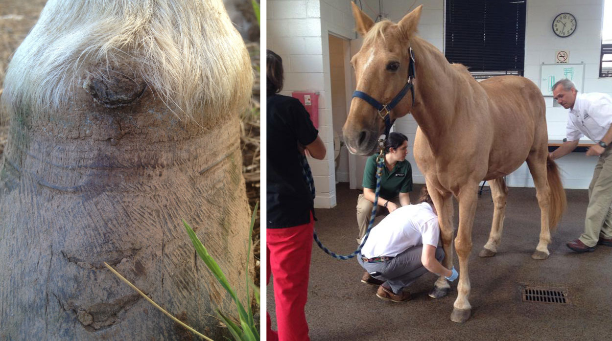
Snapper’s right foot (left photo) during his initial vet appointment (right photo) at UGA
During the lameness exam, Snapper was lame in the right front foot at a walk. At the trot, he was lame in his right front foot in addition to his right hind foot. The lameness improved after the doctors performed a basisesmoid nerve block on his right front foot.
Radiographs were taken of both of Snapper’s front feet, at multiple angles. After obtaining the images they wanted, Snapper was brought into a temporary stall and the intern brought Bridget into the radiograph room so that the doctor could go over their findings with her.
Location of Snapper’s Keratoma
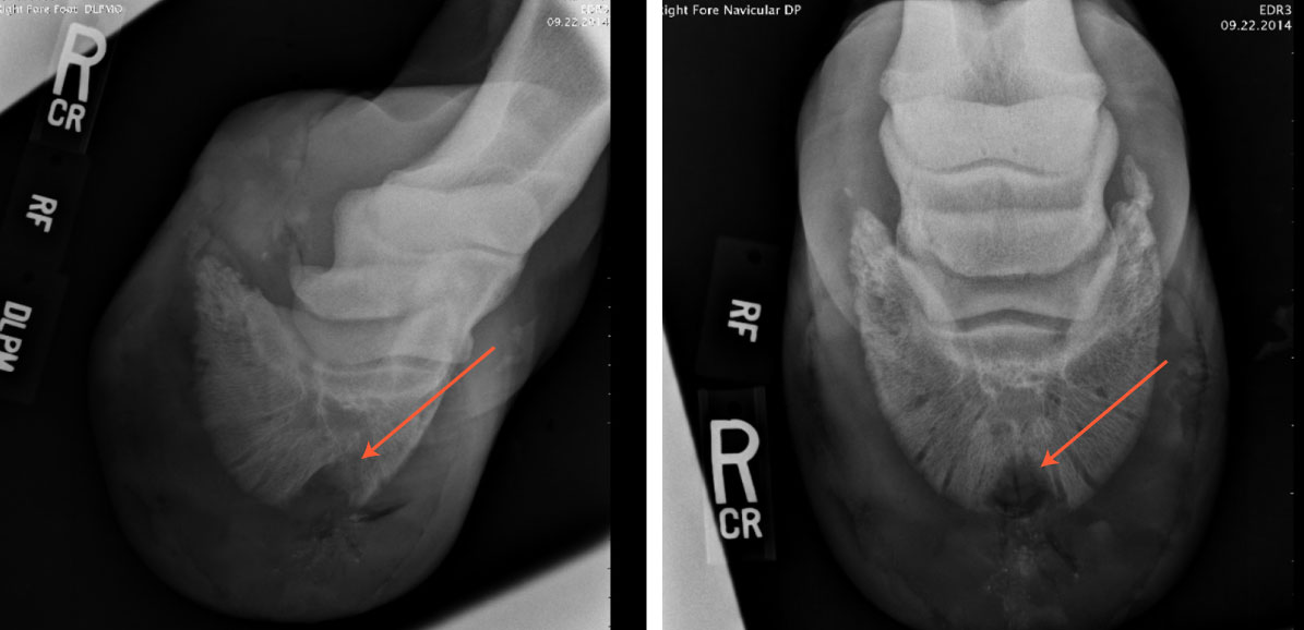
The doctor showed Bridget the radiographs of Snapper’s feet. The x-rays taken of Snapper’s left foot were normal. However, those taken of the right foot revealed the presence of an indentation in the coffin bone inside the hoof, due to loss of calcium in the bone. This indentation was likely caused by the pressure from a keratoma growing between the hoof wall and the coffin bone.
As a result of the deterioration of the coffin bone, Snapper would never again be 100% sound. The doctor recommended surgery to remove the keratoma and debride the abscess. The surgery would involve cutting a square hole into the front of Snapper’s hoof, so that they could extract the mass from his foot. Post-surgical recovery would be long as it would take up to a year for the hoof to grow back. Bridget chose to proceed with the surgery.
Surgery: Extraction of the Keratoma
Two weeks later, Snapper was brought to UGA for surgery. The procedure was performed while Snapper was standing under sedation. The doctor first removed an area of the outer hoof wall (1.5” x 1.5” square), to allow access to the tumor, which was located between the hoof wall and the deeper sensitive tissues. Bleeding was controlled with the use of a tourniquet, and the keratoma was carefully separated and removed from the hoof.
After the surgery, Snapper was hospitalized overnight for observation in case of any post-surgical complications. He was given antibiotics (penicillin) and an anti-inflammatory (bute) to help with the pain. The next day, Snapper was discharged from the hospital with detailed post-surgical care instructions for Bridget.
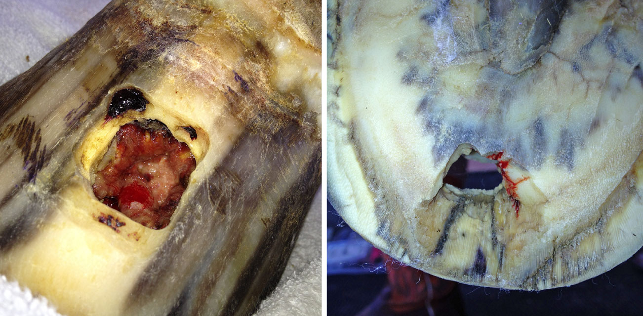
Holes in Snapper’s right front hoof wall (left photo) and on the bottom sole of his foot (right photo) from the surgery.
Post-Surgical Care
Post-surgical care of Snapper’s foot was a time-consuming process which lasted over several months, and involved stall rest, frequent bandage changes, and administering medication. It was important that Snapper’s right hoof was kept clean and dry while the hole in the hoof wall healed, for the opening in his foot made him more vulnerable to getting a bone infection. Also, since Snapper was at risk of left front support-limb laminitis, Bridget had to monitor him daily for any signs of discomfort or lameness on his left front foot.
Bandage Management: The doctors showed Bridget exactly how to bandage it before she left the hospital. She kept his foot bandaged at all times, changing it every 2 days for the first week and then every 2 to 3 days until a layer of horn formed over the hole. Bandaging Snapper’s foot was a 9-step process, which consisted of the following:
- Step 1: Remove old bandage and gauzes.
- Step 2: Use sterilized saline in a syringe to clean the hole in the hoof wall.
- Step 3: Check for any abnormalities and take photos to email to UGA.
- Step 4: Pack both the hole in the front hoof wall and on the bottom sole of the foot with amikacin-soaked gauze.
- Step 5: Cover the holes with sterile gauze.
- Step 6: Cover the hoof with sheet cotton.
- Step 7: Wrap the hoof with Vet wrap.
- Step 8: Apply duct tape boot to the bottom of the hoof.
- Step 9: Apply elastikon from the edge of the duct tape to above the fetlock.
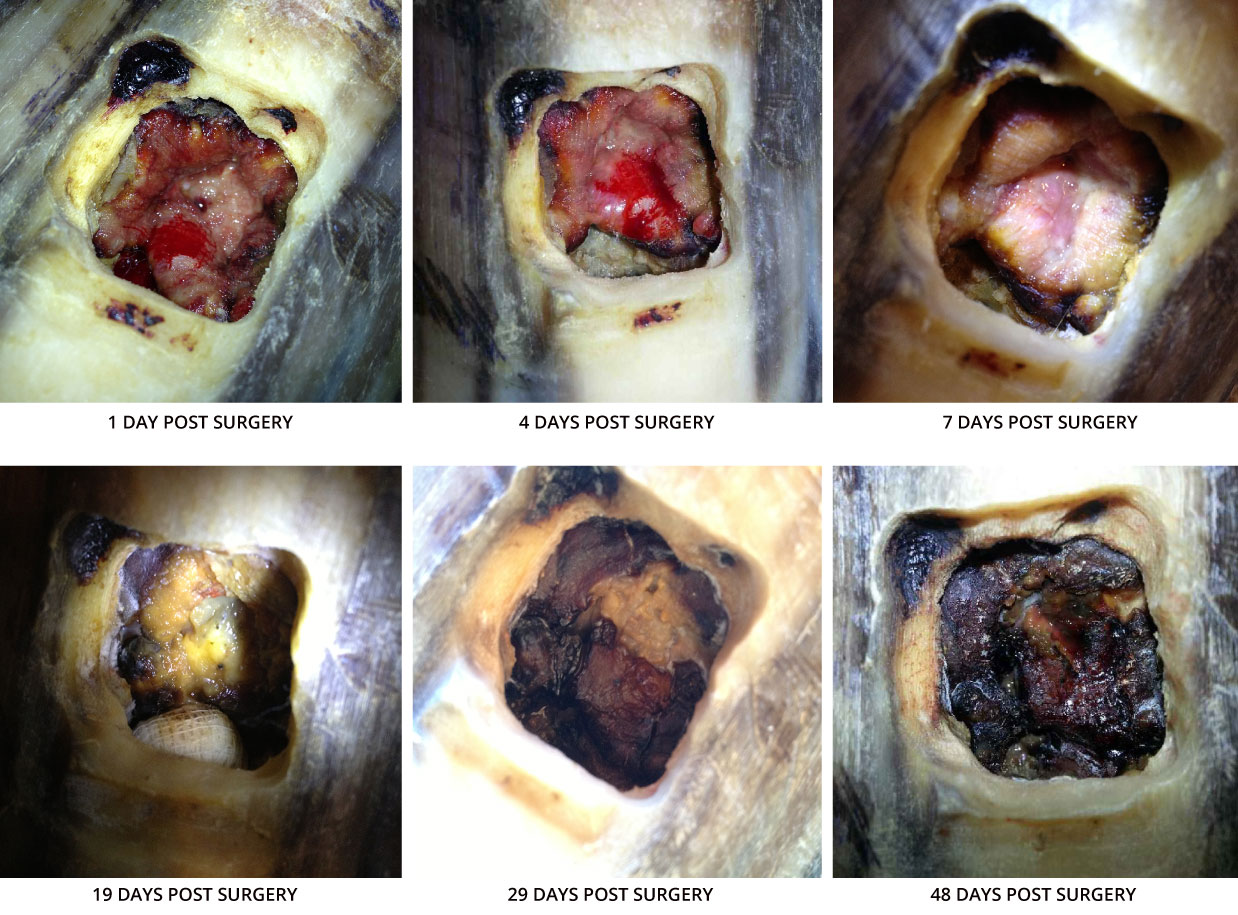
Snapper's healing timeline
Snapper received 15 tablets of Trimethoprim sulfa (960 mg tablets), dissolved in water and administered by mouth twice a day, for 10 days and bute (1 gram tablets) given by mouth twice a day for 5 days.
Oral Supplements: Bridget added a hoof supplement (Farrier’s Formula) and Karbo Combo+ (a supplement used to help boost the immune system) to his diet for several months.
Stall Rest: Snapper was kept on strict stall rest for the first two weeks following the surgery. While in his stall, Bridget had to frequently clean his stall and keep a deep layer of clean and dry shavings for him to stand one. After two weeks, as long as the paddock was dry and there was no chance of rain, Snapper was able to go out in a small paddock for a couple hours a day. His foot had to remain bandaged.
4-week Post Surgery Vet Visit
Four weeks post-surgery, Bridget brought Snapper back to UGA for a follow-up appointment. Upon arrival, the UGA veterinary team greeted them and proceeded to examine his foot. They removed Snapper’s bandage and thoroughly cleaned the holes in his right foot with chlorhexidine and rinsed with sterile saline. To help Snapper’s foot heal faster, the doctor debrided a small amount of soft yellow tissue to expose healthy, bleeding tissue. The doctors were happy with how Snapper’s foot was healing. They then repacked and bandaged Snapper’s foot and sent him home.
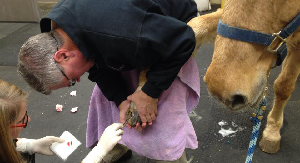
Snapper’s 4-week follow up visit at UGA
Once a healthy layer of horn formed over the hole, which took an additional four weeks, Bridget was given the okay by the doctor to allow Snapper’s foot to be left un-bandaged. Since the tissue over the hole was hard enough to block any debris---as long as it didn’t get wet. This meant that Snapper was still on controlled paddock turnout, and days where it looked like rain he had to stay inside.
8-month Post Surgery Vet Visit
It took about 8 months for his hoof to fully grow back. Bridget brought him back to UGA at this time for a follow up exam. The physical exam showed that he had small area of white line disease in his right foot, in the area of the where the hoof had grown out.
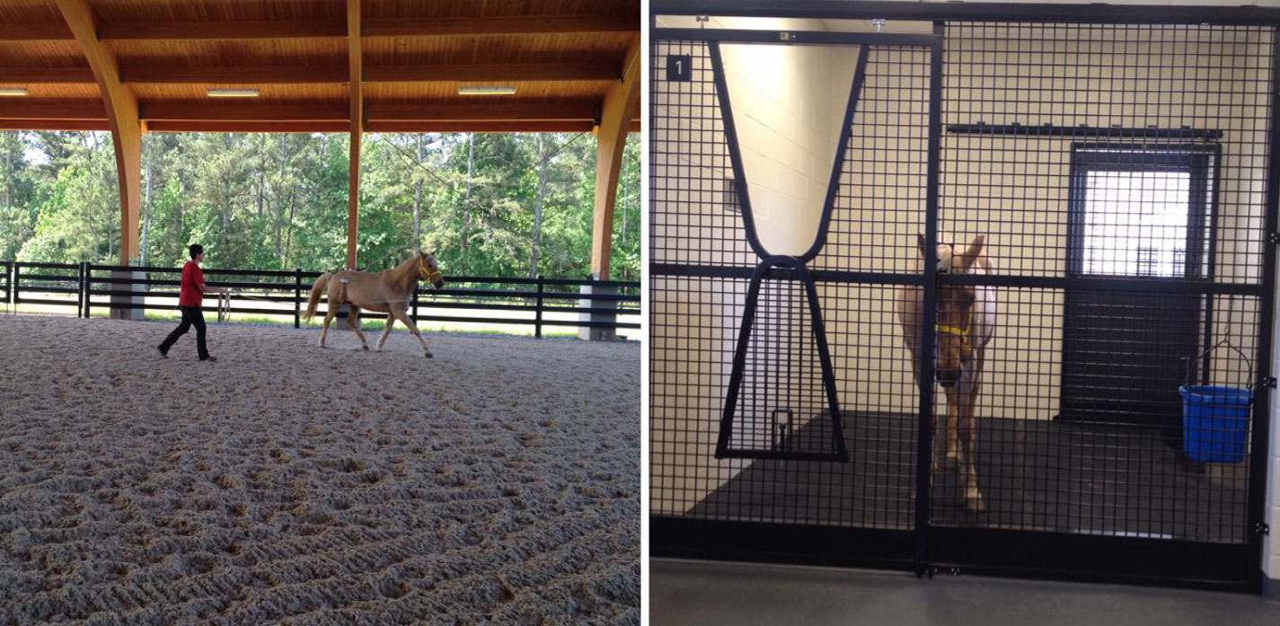
Snapper’s lameness exam (left photo) and resting (right photo) at his 8 month follow up vet appointment at UGA
During the lameness exam, Snapper was moderately lame in both of his hindlimbs and on his right front foot (grade 3 of 5). Radiographs were taken of both of Snapper’s front feet, which revealed that his left foot had developed mild ossification of the medial collateral cartilage and the right foot had ossification of both collateral cartilages, as well as mild chronic rotational laminitis.
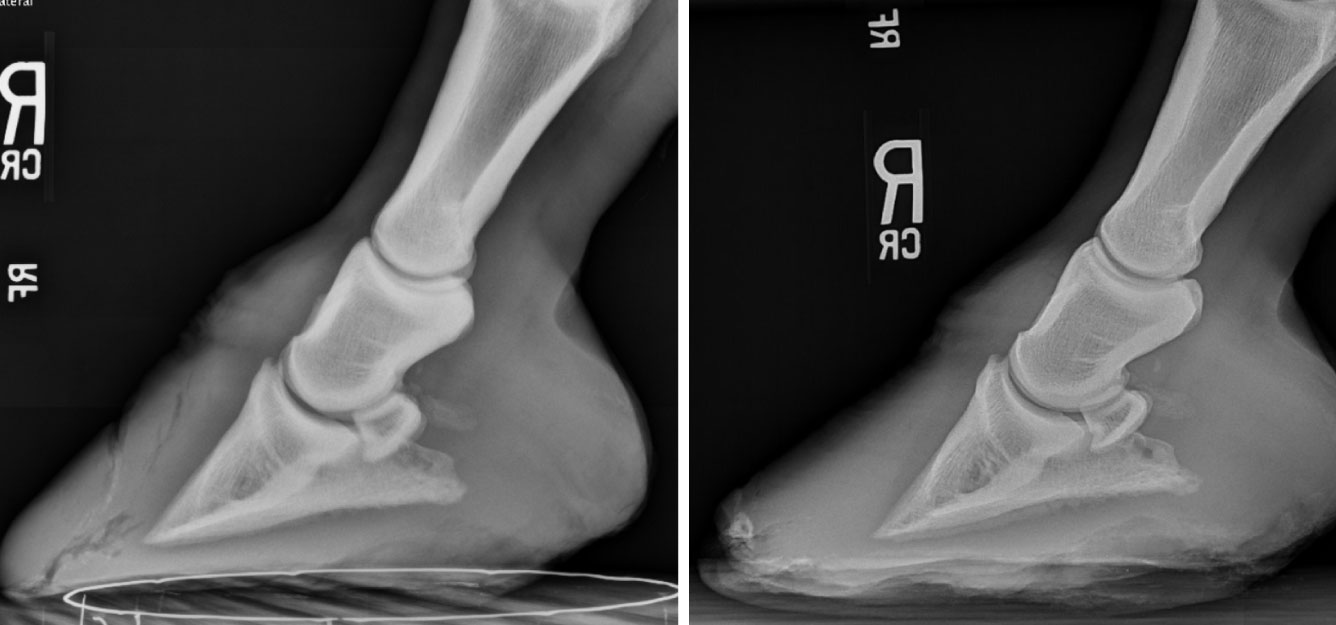
Comparison of Snapper’s right foot before surgery (left image) and 8 months after surgery (right image)
In order to help make Snapper more comfortable, due to slight rotation of his pedal bone, the doctor recommended that Snapper’s farrier use the radiographs as guidelines to help move the break over point backwards. In order to do so, he suggested the farrier to continue to shorten Snapper’s toe while concurrently raising the heel height. Being an older horse, Snapper also suffered from osteoarthritis, which is what was causing the hindlimb lameness. The doctor recommended different medications to help make Snapper more comfortable during retirement.
Bridget’s Reflections on the Experience
“UGA has an exemplary large animal clinic and team and we could not be happier with our experience there. Between our horrible colic situation and then the keratoma I would hands down recommend them to anyone. Although abscesses are common, it is not common for them to reoccur as often as Snapper's did. Something may be seriously wrong if they keep getting constant abscesses and it needs to be addressed ASAP before irreversible damage can be done. When you take your horse to the vet to get a diagnosis and you keep getting the same answer then keep moving around to other vets and seek a specialist until you find a better answer. In the medical field not all conditions are known to all veterinarians and it is important to get a second opinion.
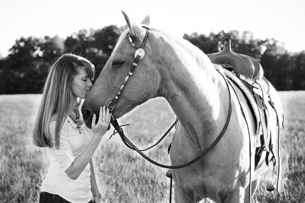
Bridget and Snapper
About Wild Hound Outfitters
Wild Hound Outfitters is a boutique pet product business which sells unique and beautiful leather dog collars and other handcrafted products for dogs and their owners. Bridget and Trevor Tuckey founded the business in 2013, where it was originally a small Etsy shop named J&B Custom Leather which originally focused primarily on dog collars. Since then, they have expanded their business and sell a wide variety of products through their website, although they still also operate through Etsy.
Follow them:
About the University of Georgia
The University of Georgia (UGA) Veterinary Teaching Hospital, located in Athens, Georgia, handles more than 25,000 cases of small and large animal visits each year and offers 25+ specialty services, including a 24-hour emergency service. Their hospital staff consists of board-certified specialists, resident veterinarians and interns, veterinary students, and technical staff. The hospital offers brand new, state-of-the-art facilities and top-of-the-line equipment such as a CT Scanner, MRI, and a linear accelerator. The campus where the Hospital is located is referred to as the Veterinary Medical Center. It includes the main Hospital, a covered equine performance arena, a building for the Hospital’s ambulatory services, and an education building for teaching third-year veterinary students.
Follow them: